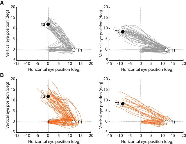
Figure 3.
Eye position traces for a representative HC (A) and SZP (B). The eye position between the initiation of the first saccade toward T1 (empty circle) and the termination of the second saccade toward T2 (filled circle) are depicted for each trial in which successive saccades to T1 and T2 were produced. Plots are separated for 90° (left) and 135° (right) T1–T2 angles. Saccade vectors to different target locations were normalized into the same space.

