Fig. 1.
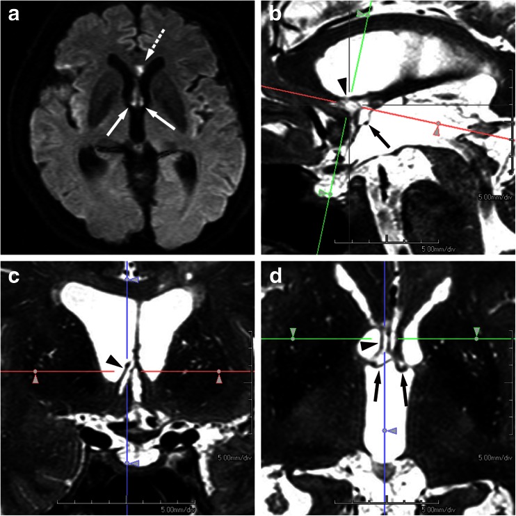
A 67-year-old man with a ruptured aneurysm of the ACoA was treated by transarterial coil embolization on the day of onset. Disorientation was suspected on the next day of embolization, but formal neuropsychological assessment was hardly valid due to confusional state in the acute phase. Diffusion-weighted image performed on the 4th day of embolization (a) demonstrates acute infarction in the column of fornix, bilaterally (arrows) and in the rostrum of the corpus callosum (dashed arrow). Two months later, formal neuropsychological tests revealed amnesia, confabulation, and disorientation. Paramedian sagittal (b), coronal (c), and axial (d) MPR images were generated from 3D data of T2-weighted volumetric isotropic turbo spin-echo acquisition (VISTA). The coronal image (c) corresponds to the green line in the paramedian sagittal (b), and the axial image (d) corresponds to the red line in the paramedian sagittal (b), so that both images pass through the superior part of the paraterminal gyrus in the right (arrowheads in b–d). Infarcted foci involve not only the pars libera of the column of the fornix bilaterally (arrows in d) and the rostrum of the corpus callosum (not shown) but also the paraterminal gyrus in the right and the anterior commissure (arrow in b). Thus, it may well be said that the involvement of the paraterminal gyrus and the anterior commissure found on 3D images in the chronic phase had not been visualized on the initial 2D diffusion-weighted images in the acute phase

