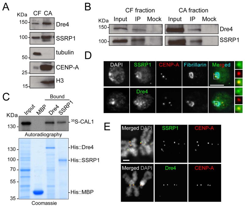
Figure 3. FACT interacts with CAL1 and localizes to the centromere in S2 cells.
A) Western blots of chromatin-free and (CF) chromatin-associated (CA) extracts from S2 cells with indicated antibodies. Tubulin and histone H3 antibodies are positive controls for their respective fractions. B) Western blots of IPs with anti-CAL1 antibodies from CF and CA extracts. Mock are IPs with rabbit IgGs. C) Direct interaction between in vitro translated 35S-methionine-labeled CAL1 with recombinant His::Dre4 or His::SSRP1 bound to Ni-NTA beads. His::MBP, negative control. D) IF with anti-SSRP1 or anti-Dre4 (green), anti-CENP-A (red), and anti-fibrillarin (blue) antibodies. DAPI shown in gray. Insets show 3× magnifications of boxed centromere. Bar 5μm. E) IF on metaphase chromosomes with anti-SSRP1 or anti-Dre4 (green), and anti-CENP-A (red) antibodies. DAPI shown in gray. Bar 1μm. See also Figure S2 and Table S1.

