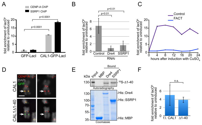
Figure 4. FACT is required for CENP-A deposition-coupled transcription.
A) CENP-A and SSRP1 ChIP-qPCR in CAL1-GFP-LacI and GFP-LacI lacO cells. The graph shows the enrichment of induced (24h) relative to uninduced cells. Error bars, 95% CI of 3 technical replicates. Significant p-values (unpaired t-test) are shown. B) qRT-PCR lacOb transcripts in CAL1-GFP-LacI cells induced (24h) 6 days after the indicated RNAi treatments. p-values (unpaired t-test) are shown. C) qRT-PCR lacOb transcripts in control (purple) and SSRP1/Dre4 RNAi (blue) cells at the indicated times. Error bars, SD of 3 technical replicates. D) IF with anti-CENP-A (red) and anti-GFP (green) antibodies in lacO cells expressing full length CAL1-GFP-LacI (top) or CAL1Δ1-40-GFP-LacI (bottom). DAPI is shown in gray. Arrow points to the lacO site. Bar 1μm. E) Direct interaction between in vitro translated 35S-methionine-labelled CAL1Δ1-40 (35S-Δ1-40) with recombinant His::Dre4 (Dre4) or His::SSRP1 (SSRP1) bound to Ni-NTA beads. His::MBP (MBP) is a negative control. F) qRT-PCR of lacOb transcripts in induced cells (24h) transiently expressing full length (f.l.) CAL1-GFP-LacI or CAL1Δ1-40-GFP-LacI. Shown is mean ±SEM of 3 experiments p=0.68 (not significant; unpaired t-test).

