Figure 1.
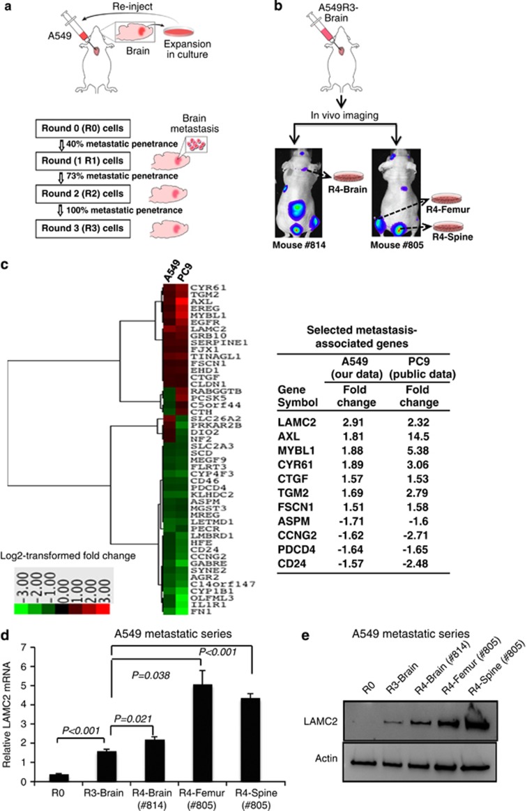
Identification of LAMC2 as a putative metastasis marker of NSCLC. (a) A schematic illustration of the in vivo mouse metastasis model with repeated intracardiac injections of A549 cells. Outgrowth of A549 cells from the cultured brain tissue indicated that injected A549 cells metastasized to the brain. A549R1, A549R2, and A549R3 cells were serially established by performing intracardiac injection of A549R0 (5 × 10⁵ cells), A549R1 (2.5 × 10⁵ cells), and A549R2 (2.5 × 10⁵ cells), respectively. For each round, 10 mice were injected. (b) Bioluminescent images after intracardiac injection of luciferase-transfected A549R3-Brain cells, demonstrating non-brain tropism. A549R4-Brain, -Femur, and -Spine cells were established after culturing metastatic cells derived from the corresponding organ metastases after injection of A549R3 cells. (c) Expression profiles of A549 (our data) and PC9 metastatic cells (public data, GSE14107), which were independently derived using the intracardiac injection mouse metastasis model, revealed 48 differentially expressed genes between R0 and R3 that were common in both systems, with criteria of false discovery rate <1.0 and log 2 fold change >1.5 or <−1.5. Differentially expressed genes were further narrowed down to 11 based on the unidirectional log 2 fold change across R1, R2, and R3, and including only genes reported to have cancer-associated function. (d) Real-time qRT-PCR analysis of LAMC2 in A549R0, A549R3, and A549R4 cells. Student's t-test was performed to compare A549R0 versus A549R3 and A549R3 versus A549R4. Data are presented as mean±S.E.M. of triplicate experiments. (e) Immunoblot analysis of LAMC2 in A549R0, A549R3, and A549R4 cells

