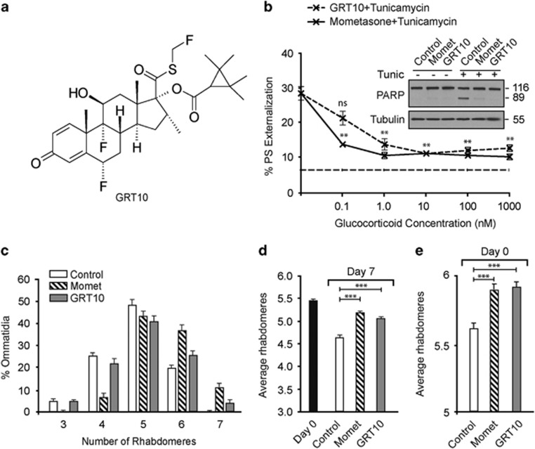
Figure 3.
The transrepression arm of glucocorticoid signaling is sufficient to protect against ER stress-induced apoptosis and HTT-mediated neurodegeneration. (a) Chemical structure of GRT10. The transrepression and transactivation assays characterizing the specificity of GRT10 have previously been published, where GRT10 is denoted as compound 5.29 (b) HeLa cells, exposed for 4 h to the indicated concentrations of GRT10 and mometasone, followed by a further 20 h of tunicamycin (10 μM), were analyzed by FACS for PS externalization (n=3). Dashed, straight line at the bottom depicts the extent of basal cell death in control cells. Statistical analysis was conducted using a paired t-test (**P<0.01, NS, not significant, if P>0.05). The inset shows the extent of apoptosis assessed by cleavage of PARP. (c) Newly emerged fruit flies, expressing mutant HTT, were exposed to either mometasone (10 μM, n=14) or GRT10 (10 μM, n=11) for 7 days and the number of rhabdomeres per ommatidium was scored by pseudopupil analysis. (d) Same as (c), but the average number of rhabdomeres per ommatidium has been plotted. Statistical analysis was conducted by analysis of variance (ANOVA) with Newman–Keuls post hoc tests (***P<0.001). (e) Same as (d), but the analysis was carried out with day 0 flies to assess HTT-mediated neurodegeneration during development (n=11–13). Statistical analysis was conducted by ANOVA with Newman–Keuls post hoc tests (***P<0.001)

