Figure 4.
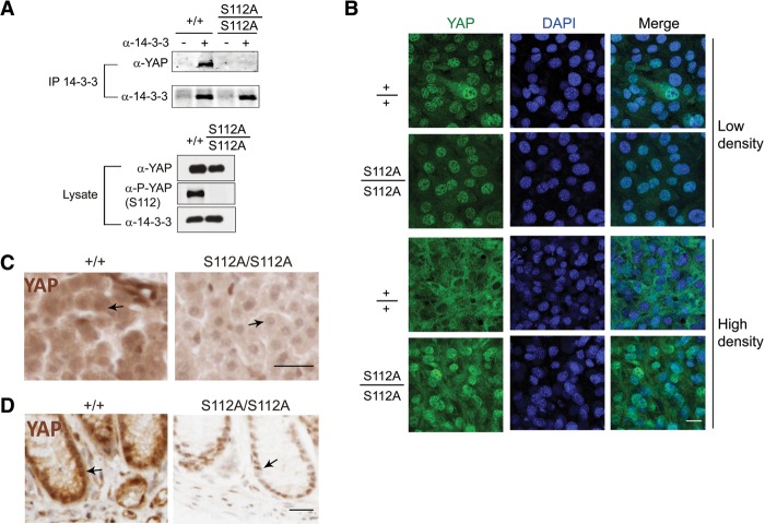
S112 phosphorylation is required for 14-3-3 binding and cytoplasmic translocation of endogenous YAP induced by contact inhibition in cell culture or developmental Hippo signaling in intact tissues. (A) Coimmunoprecipitation assay. Cell lysates of wild-type and YapS112A/S112A MEFs were immunoprecipitated with α-14-3-3 antibody and immunoblotted with α-YAP antibody. (B) Loss of cell contact-induced YAP translocation in the YapS112A/S112A MEFs. Wild-type and YapS112A/S112A MEFs grown at low or high density were immunostained for endogenous YAP (green) and nuclear dye DAPI (blue). Endogenous YAP shows nuclear-to-cytoplasmic translocation at high cell density in the wild-type cells but not in the YapS112A/S112A cells. Bar, 50 μm. (C) Immunostaining of YAP in liver sections from wild-type and YapS112A mice. Note the more prominent nuclear localization of YAP in the YapS112A liver compared with the wild-type liver (arrows). Also note the overall decrease of YAP staining intensity in the YapS112A liver. Tissue sections were processed in parallel and stained under identical conditions. Bar, 50 μm. (D) Immunostaining of YAP in colon sections from wild-type and YapS112A mice. Note the decrease of overall YAP staining and the more prominent nuclear localization in the colonic epithelial cells in YapS112A mice compared with the wild-type mice (arrows). Tissue sections were processed and stained under identical conditions. Bar, 50 μm.

