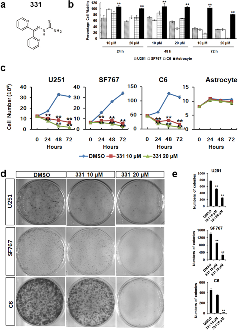
Figure 1. Compound 331 selectively induced cell death in glioma cells.
(a) The structure of compound 331. (b) Compound 331 treatment induced cell death in human glioma cells (U251, SF767) and rat glioma cells (C6) but not in normal rat astrocytes. All cells were treated with compound 331 at 10 μM or 20 μM for 24 h, 48 h and 72 h. Data represents the mean ± S.E.M. of three independent experiments. **p < 0.01 compared with glioma cells. (c) Compound 331 treatment induced significant proliferation inhibition in human glioma cells (U251, SF767) and rat glioma cells (C6) but not in normal rat astrocytes. All cells were treated with compound 331 at 10 μM or 20 μM for 24 h, 48 h and 72 h. Data represents the mean ± S.E.M. of three independent experiments. **p < 0.01 compared with control. (d) Glioma cells were treated with DMSO as control or compound 331 (10 μM, 24 h or 20 μM, 24 h). The cells were then collected and 3000 cells were incubated in medium for an additional 10 days. Cells were then stained with crystal violet (0.4 g/L). (e) The numbers of colonies per dish. Data represents the mean ± S.E.M. of three independent experiments. **p < 0.01 compared with the controls (DMSO).

