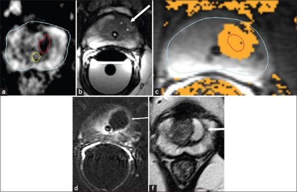
Figure 2.

Magnetic resonance-guided focal laser ablation treatment. A 54-year-old male with biopsy-confirmed Gleason 7 (3+4) prostate carcinoma. (a) Pre-treatment axial apparent diffusion coefficient map (ADC) shows a well-demarcated magnetic resonance imaging (MRI) visible lesion (outlined in red) in the left transition zone at the level of mid gland. The prostatic urethra is outlined in yellow. (b) Intra-operative axial balanced steady-state precession sequence MRI scan confirming final position of two transperineally advanced cannulas with gadolinium markers (arrow) prior to initiating power. (c) Thermal map image during treatment showing areas of heat deposition color coded (orange) overlaid on tumor outline (in red). (d) Immediate post-treatment axial post-contrast Gd-DTPA (Magnevist®, Bayer Healthcare) enhanced subtraction image highlights the devascularized ablated volume (arrow), showing no damage to the rectal mucosa or neurovascular bundle or even the adjacent urethra, which is outlined by Foleys catheter. (e) Post-treatment axial T2-weighted image at 6 months shows devascularized cystic area at the site of treatment. All six samples obtained from the site at the 6-month follow-up biopsy were negative
