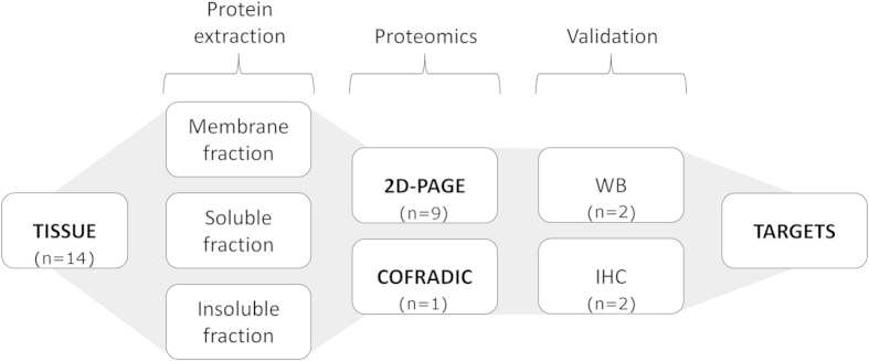
Figure 1. Schematic representation of the experimental procedures performed.

Three tissue portions were isolated from rat brains at 48 h post-ischemia: infarcted tissue (lesion), as delimited by TTC staining; a 2-mm strip around the infarct region (peri-infarct); and control tissue from the contra-lateral hemisphere. The protein content from each portion was extracted in three different fractions (membrane, soluble and insoluble). The samples were analysed 2D-PAGE and COFRADIC proteomics. Proteins with significant differences of expression between the tissue portions (>3-fold, p < 0.05) were further studied by western blotting (WB) and immunohistochemistry (IHC). Data were combined to define suitable molecular targets of the peri-infarct tissue.
