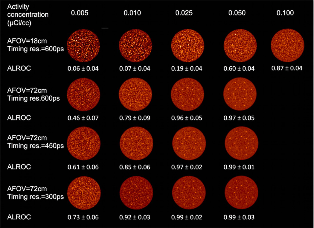
Figure 2.
Reconstructed images of the central transverse slice from the lesion phantom. Images are from a single data replicate and are shown for varying activity concentration (columns), and combination of AFOV and timing resolution. The lesions are 1 cm in diameter with 3:1 uptake relative to the background.

