Figure 2.

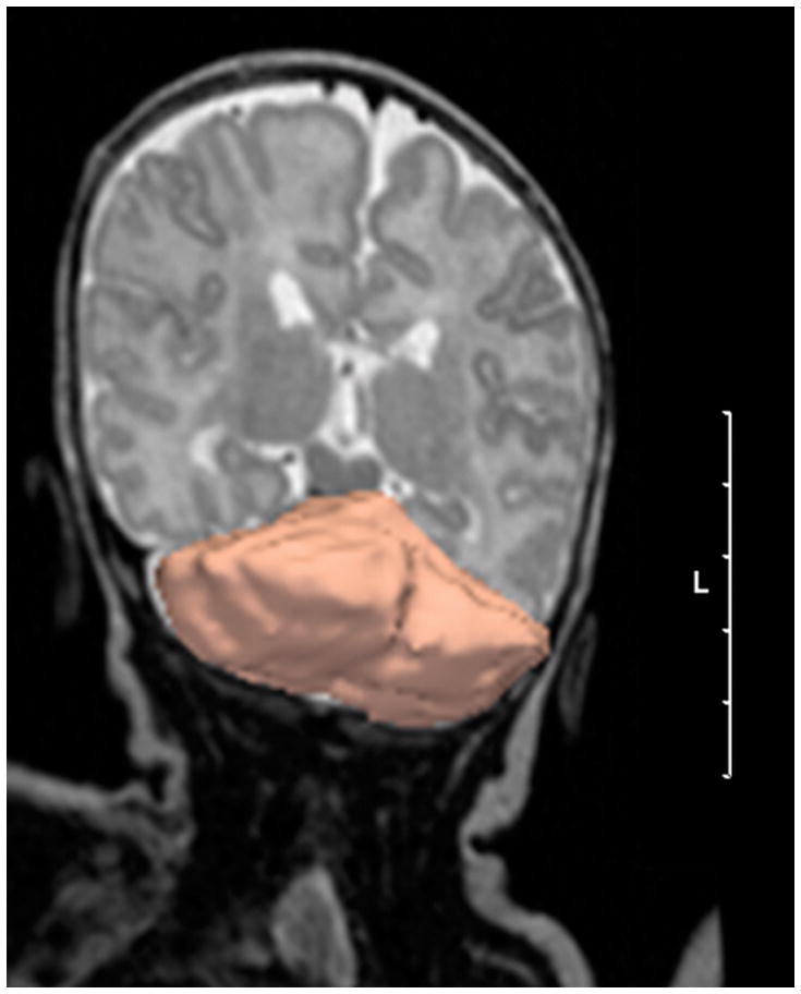
3D reconstruction of manual cerebellar outlining of reconstructed 3D image slices using 3D Slicer software. (Scale bar = 5 cm, L = left). The figure is distorted to give a 3D impression of the cerebellum.

3D reconstruction of manual cerebellar outlining of reconstructed 3D image slices using 3D Slicer software. (Scale bar = 5 cm, L = left). The figure is distorted to give a 3D impression of the cerebellum.