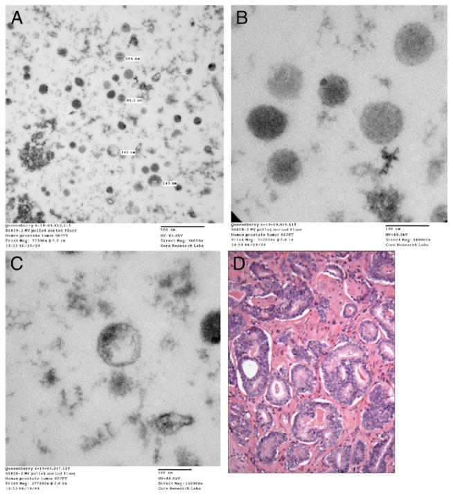
Figure 3.
Transmission electron microscopy reveals microvesicles from prostate tumor samples cultured without BM cells for 7 days. CM was removed and microvesicles were isolated by high speed centrifugation. Sections were examined by transmission electron microscope and images were collected with charge coupled device camera. A, microvesicles. Reduced from ×36,000. B, microvesicles. Reduced from ×180,000. C, microvesicles. Reduced from ×140,000. D, pathology slide shows prostate tumor cells from same patient. H & E, reduced from ×600.

