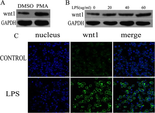
Fig. 1.

wnt1 was increased in LPS-stimulated THP-1 cells. a wnt1 protein level was increased in THP-1 cells treated with PMA (100 nmol/ml for 24 h) (p < 0.05). b wnt1 protein level was increased in a dose-dependent manner in THP-1 cells treated with LPS (0–60 μg/ml) (p < 0.05). c THP-1 cells were cultured in the presence or absence of LPS (40ug/ml) for 24 h. Confocal microscopy imaging of wnt1 (green) was shown. Results were normalized against levels of GAPDH protein.
