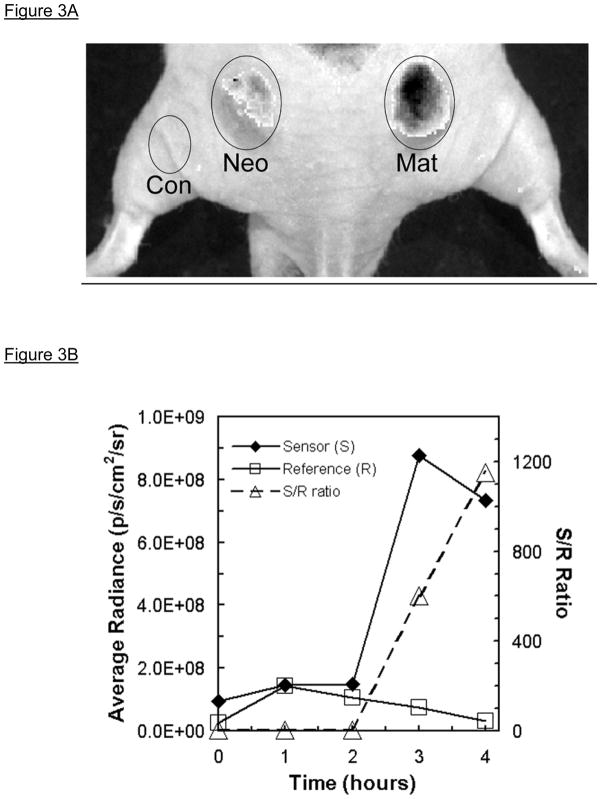
Figure 3.
Quantitative in vivo fluorescence imaging of tumor-associated MMP7 activity in mouse subcutaneous xenograft colon tumors [modified from (5)]. Panel A. Subcutaneous injection into a nude mouse of either SW480neo (Neo) or SW480mat (Mat) colon tumor cells resulted in xenograft tumors (left and right, resp.) within 3–4weeks; the mouse (dorsal, caudal view) was imaged in the Cy5.5 Sensor (S) channel 4hr post retro-orbital intravenous injection of PB-M7NIR (white light photograph overlaid with fluorescence image shown in gray scale). There is significant sensor signal associated with the SW480mat tumor indicative of cleavage of PB-M7NIR by MMP-7 with minimal signal in the SW480neo control tumor that is similar to that over a non-tumor control (Con) region. Panel B. Sensor (S) and reference (R) signals measured in an SW480mat tumor reveal increasing signal in the sensor channel over time. The S/R ratio increases markedly after 2h as the uncleaved reagent clears the circulatory system.

