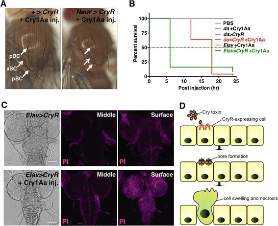
Fig. 7.

In vivo cell ablation of peripheral or central nervous system by Cry1Aa injection. a Adult flies injected with Cry1Aa during third instar larval stage lost their bristles. Arrows in the left panel (negative control, without Neur-Gal4) indicate bristle positions (pDC, posterior Dorsocentral; aSC, anterior Scuteller; pSC, posteior Scuteller) and arrows in the right panel (Neur > CryR) indicate the presence (aSC) or absence (pDC, pSC) of bristles. b Survival curve of male flies with CryR expression throughout the whole body (da) or only in neurons (Elav) with and without Cry1Aa; n = 50 for each condition. c Confocal images of Propidium iodide (PI) staining of larval brain from Elav > CryR, with and without Cry1Aa injection. Brains were dissected 3 h post-injection and then stained with PI. A single focal plane from the middle and surface regions are shown. d Schematic view of selective cell ablation by the Cry1Aa/CryR system. Cry1Aa specifically induces cell swelling and necrosis in CryR-expressing cells by pore formation in the plasma membrane
