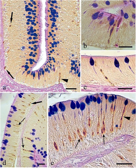
Fig. 6.

Histological sections from the intestines of Squalius cephalus infected with the acanthocephalan Pomphorhynchus laevis and treated with either the galanin (a-c) or the serotonin (d-e) antisera and a subsequent AB/PAS stain for mucous cells. There are numerous endocrine cells that are immunoreactive to the two antisera; many of these are close to the goblets of the mucous cells. The immunoreactive endocrine cells also display a range of differing morphologies including: the closed type (a, d, thick arrows), the open type with a mid-epithelial body (d-e, thin arrows), and, the open type with a “reservoir-like” appearance to them (a-c, e, arrowheads). Scale bars 20 μm
