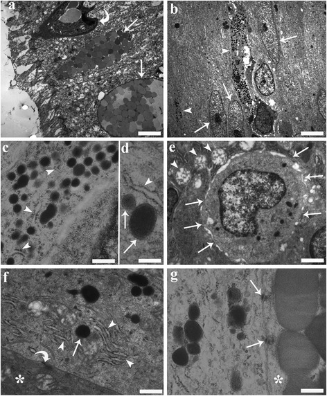
Fig. 9.

Transmission electron micrographs from the mid-gut of a specimen of Squalius cephalus infected with Pomphorhynchus laevis. a Two mucous cells (arrows) containing numerous mucus granules of various size and electron densities that are positioned close to the surface of the epithelium. The curved arrow highlights the presence of a rodlet cell. b Two endocrine cells (arrow heads) with long, narrow extensions directed toward the lumen are seen positioned between gut enterocytes (arrows). c Numerous secretory granules within the cytoplasm of an endocrine cell that is in close proximity to rough endoplasmic reticulum (RER) (arrow heads). d A fine electron-dense material can be seen filling the inner part of the granules (arrows); an electron-lucent halo separates the core of the granule from the membrane; the relationship between the RER (arrow head) and granules is visible. e The electron micrograph shows the basal portion of an endocrine cell that contains an euchromatinic nucleus and has numerous connections (arrows) with the surrounding mucous cells. Note the round mitochondria (arrow heads) with moderate cristae within the mucous cells. f An endocrine cell adjacent to a mucous cell (asterisk), with well-developed RER (arrow heads) close to a secretory granule (arrow). Also note the different electron-densities of the cytoplasm of the two cells and the presence of a desmosome (curved arrow). g An endocrine cell connected to a mucous cell (asterisk) by two desmosomes (arrows). Scale bars: a: 3.14 μm; b: 3.57 μm; c: 0.39 μm; d: 0.25 μm; e: 1.06 μm; f: 0.55 μm; g: 0.33 μm
