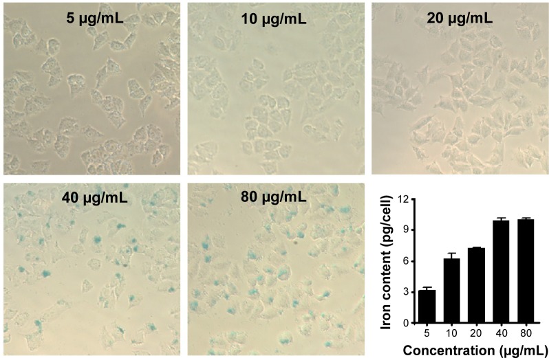
Figure 10.
Microscope images of Prussian blue dye staining of MCF-7 cells after 24 hours of incubation at concentrations of 5 μg/mL, 10 μg/mL, 20 μg/mL, 40 μg/mL and 80 μg/mL, respectively.
Notes: The iron in the cells was stained blue and the Fe–iron accumulation in MCF-7 cells was detected by ICP-OES analysis shown in the bottom-right image.
Abbreviation: ICP-OES, inductively coupled plasma atomic emission spectroscopy.

