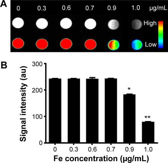
Figure 12.

T2-weighted MR images and quantitative signal intensity analysis of Fe3O4@SiO2/PEI/VEGF shRNA.
Notes: (A) T2-weighted MR images and color T2-weighted MR images of Fe3O4@SiO2/PEI/VEGF shRNA. The weight ratio of Fe3O4@SiO2/PEI to VEGF shRNA is 40:1. (The color bar from red to blue shows the gradual decrease of MR signal intensity.) (B) Quantitative signal intensity analysis. *P<0.05 and **P<0.01 compared with the control (0 μg/mL).
Abbreviations: MR, magnetic resonance; PEI, polyethylenimine; shRNA, small hairpin RNA.
