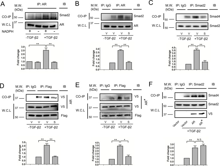
Figure 6.
Aldose reductase inhibitors inhibit AR-SMAD2 and SMAD2-SMAD4 interactions. The HLE B3 cells were treated with or without TGF-β2 (5 ng/mL) in the presence or absence of NADPH during co-immunoprecipitations (A).The HLE B3 cells were pretreated with vehicle (V) or Sorbinil (S, 10 μM) for 1 hour followed by TGF-β2 (5 ng/mL) for 1 hour (B–E). Co-immunoprecipitation was performed by using AR or SMAD2 antibody for pull down and SMAD2 (B) or SMAD4 (C) was detected by Western blot after pull-down assay. Immunoglobulin G was used for negative control. The SMAD2 and AR in whole-cell lysate (WCL) were used for loading control. Aldose reductase catalysis is irrelevant for SMAD2 interactions (D, E). Human lens epithelial B3 cells were co-transfected with either AR-V5 (D) or ARY48F-V5 (E) and Flag-SMAD2 plasmids followed by co-immunoprecipitation using Flag antibody for pull down. Western blot detected V5 after pull-down assay. Immunoglobulin G was used for negative control. Flag and V5 in WCL was used for loading control. Values for relative band intensity are given below the relevant sets of blots. Overexpression of AR or ARY48F (AR*) enhances TGF-β2–induced SMAD2-SMAD4 interactions (F). Changes in the level of proteins of interest were analyzed for statistical significance and are displayed in graphical form as mean values ± SEM (n = 3). *P < 0.05; **P < 0.01.

