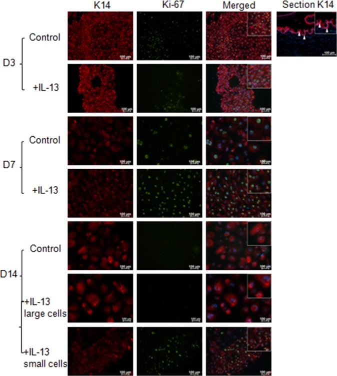
Figure 4.
K14 and Ki-67 immunostaining cultured conjuntival goblet cells. The increased proliferation with IL-13 stimulation at D3 and D7 detected by the WST assay was confirmed at the protein level with K14 (red) and Ki-67 (green) staining. At D14, the majority of the cultured cells were Ki-67 negative; however, in approximately 25% of the cultures a mixture of large, Ki-67 negative and small, Ki-67 positive cells were observed. Arrows, K14 epithelial staining in conjunctival tissue section.

