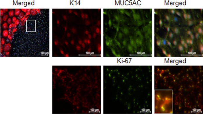
Figure 5.
Higher magnification of small proliferating cells noted in the D14 IL-13–treated conjunctival goblet cell cultures (area surrounded by white box in upper left). Staining illustrates that these small cells are K14, MUC5AC, and KI-67 positive. Inset in bottom panel shows higher magnification of K14 and Ki-67 and dual positive cells. Hoechst (DNA stain), blue; K14, red; Ki-67, or MUC5AC, green.

