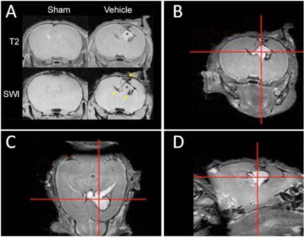
Fig. (1). CSF accumulation and ventriculomegaly 7 days after neonatal brain hemorrhage induction depicted by imaging.
Magnetic resonance imaging (MRI) was used to evaluate injury progression in sham and vehicle P14 rat pups 7 days after NBH induction. These images suggest hydrocephalus development commences as early as 1 week following NBH induction. (A) T2-weighted MRI (top row) depicts CSF accumulation (hyperintense T2-signal marked with an asterisk) and ventriculomegaly. Susceptibility weighted imaging SWI (bottom row) depicts clots within the periventricular region (hypointense signal marked with arrows). Sham animals did not have any visible brain injury. T2-weighted MRI depicting (B) coronal, (C) axial and (D) sagittal cross-sections of NBH animal depicting CSF accumulation, ventriculomegaly, and minor midline shift.

