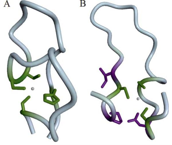
Figure 7. Binding site prediction examples.
Residues are colored as “correctly predicted” (true positive, green) and “wrongly predicted” (false positive, violet). (A) The Zn2+ binding site in a Btk-type zinc finger in the PH domain from the Tyrosine-protein kinase Tec (T0657) is formed by His 121, Cys 132, Cys 133, and Cys 143. Coloring according to prediction by group “Binding_Kihara” (FN231). (B) Structure of a hypothetical protein T0659 with a Zn2+ ion bound by three conserved Cysteine residues (Cys 43,48,63). Coloring according to predictions by group “SP-ALIGN” (FN326).

