
Figure 2.
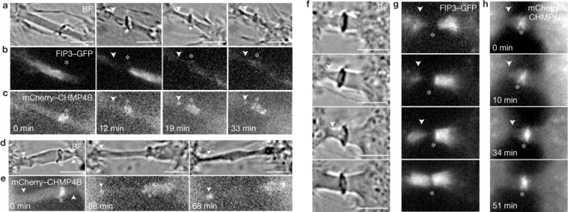
The secondary ingression forms before the recruitment of CHMP4B. Mitotic HeLa FIP3–GFP cells expressing mCherry–CHMP4B were imaged using time-lapse microscopy. (a–e) Sequential time-lapse images show abscission occurring either close (<2μm; a–c) or far away (>6μm) from the midbody (d–e). (f–h) Sequential time-lapse images show failed secondary ingression. In all images, the arrowheads mark secondary ingression and abscission sites. The asterisks mark the midbodies. Scale bars, 5 μm.