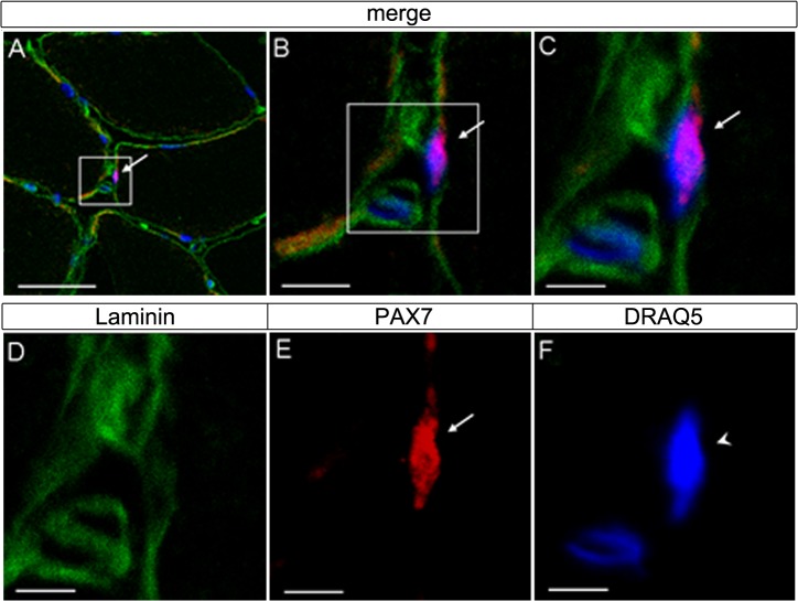
Fig 2. Immunostaining of serial cryocut cross-sections in vastus lateralis muscle of cyclist after the pre-competitive season.
(A) Muscle fibers are shown, where one area is viewed at a higher magnification (white box) in (B) and (C); (D, E, F) co-immunolocalization of Laminin (green), PAX7 (red) and myonuclei (arrowhead) counterstained with DRAQ5 (blue); the marked area in (A-C) represents the same area as shown in (D–F); SCs (arrows) are indicated. (C) Note that PAX7 positive SC is located between the sarcolemma and the basal lamina of the muscle fibre. Bars: 50 μm (A), 10 μm (B) and 5 μm (C–F).

