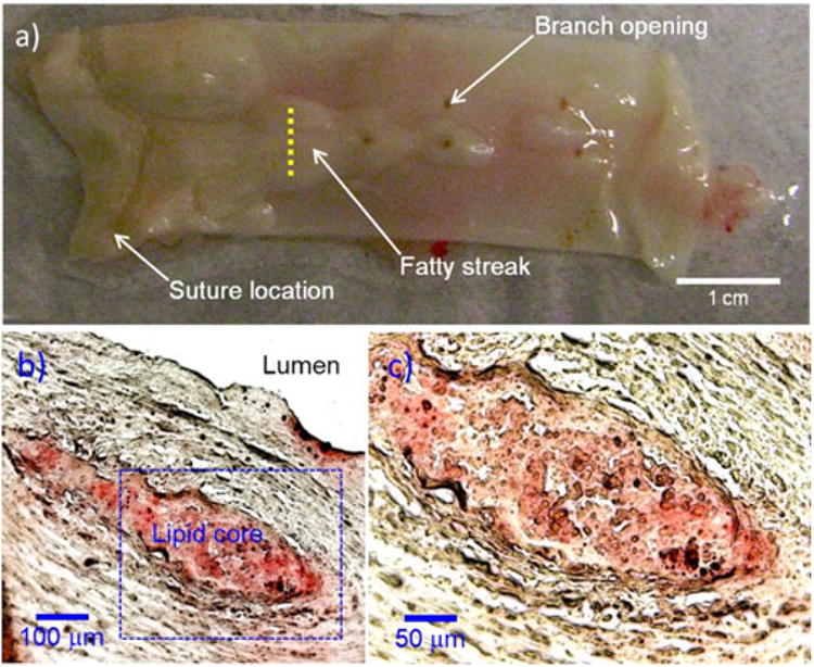
Fig 4.

Representative aorta specimen from a hyperlipidemic rabbit: a photograph of inner luminal wall, b Oil-red O-stained tissue cross section, c magnified image of the dotted blue box in (b). Histological Oil-red O stains the lipid core. The histological staining (b, c) along the yellow dashed line (a) reveals the presence of atherosclerotic plaque with lipid core.
