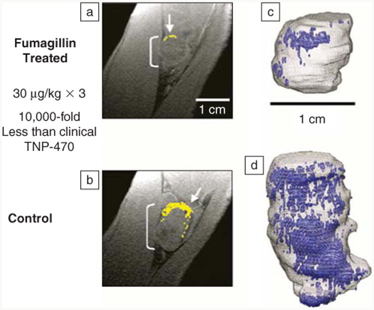
Figure 5.

(a) Diminished αVβ3-integrin contrast enhancement in a T1-weighted 3D gradient echo magnetic resonance (1.5 T) single slice image of a VX2 rabbit tumor following αVβ3-targeted fumagillin nanoparticles versus (b) those given αVβ3-targeted nanoparticles without drug. (Enhancing pixels are color coded in yellow.) (c) 3D angiogenesis maps in VX2 tumors following αVβ3-targeted fumagillin nanoparticles versus (d) αVβ3-nanoparticles without drug.36
