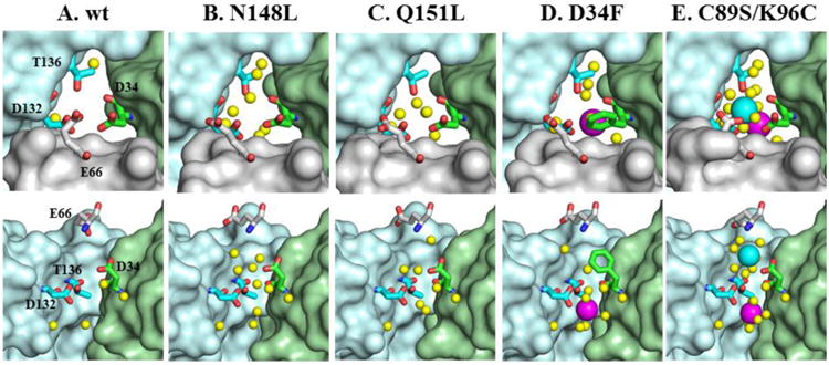
Figure 6.

Zoomed-in view of B-fold pores in wt BfrB and mutants. The top row depicts the pores viewed from the protein exterior and the bottom row shows cross-sectional views with the wheat subunit omitted. FeB-1 iron ions are shown as magenta spheres, the FeB-2 iron ion is shown as a cyan sphere, water molecules as yellow spheres, nitrogen in blue and oxygen in red.
