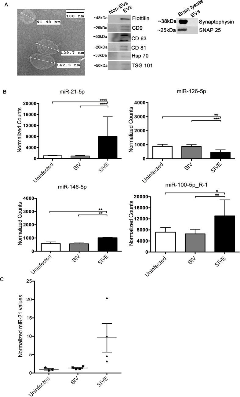
Fig 1. Isolation and characterization of brain derived EVs.
(A) Left, Electron microscopic (TEM) morphological analysis of EVs derived from uninfected (control) macaque brain. EVs show a size range from 100–150 nm. Scale bar = 100 nm. Right, Western blots for flotillin, CD9, CD63, CD81, HSP70, TSG101 markers for EVs. Non-EV fractions from sucrose gradients were used as negative controls for the EV proteins, brain lysates were used as positive controls for the synaptic proteins. (B) Small RNA sequencing performed on RNA isolated from uninfected, SIV and SIVE brains. Analysis revealed significantly increased expression of miR-21-5p, miR-100-5p and miR-146-5p, and decreased expression miR-126-5p, in SIVE. Error bars = SEM; * P <0.05, ** P <0.01*** P <0.001, **** P < 0.0001; ANOVA with Tukey’s post-hoc test. (C) qRT-PCR validation of miR-21 expression in EVs. Relative quantification was performed based on a standard curve. Statistical analyses were performed on log-transformed values. One-way ANOVA showed p = 0.0024 with Tukey’s <0.01 for uninfected vs. SIVE, and SIV vs. SIVE.

