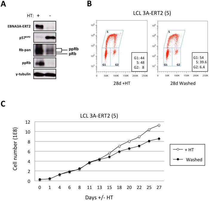
Fig 12. The increase of p57KIP2 level is associated with de-phosphorylation of Rb and reduced entry into S phase and proliferation.
(A) Western blot analysis of extracts from LCL EBNA3A-ERT2 (line 5) cultured with (+) or without (-) 4HT for 30 days showing that after inactivation (and degradation) of the EBNA3A-ERT2 fusion protein, p57KIP2 expression increases, and the amount of hyperphosphorylated Rb (ppRb) is dramatically reduced and is no longer detected using a phospho-Rb-specific antibody. The blot was probed for γ-tubulin as a control for loading. (B) Cell cycle distribution of LCL EBNA3A-ERT2 (line 5) culture with or without 4HT for four weeks was determined by flow cytometry following exposure to EdU for 2 hours (2h pulse). (C) A comparison of the population growth rate between these cells cultured with or without 4HT was analysed by counting the number of viable cells every 2–3 days. Total cell numbers were plotted at each time point. Data are representative of two independent experiments.

