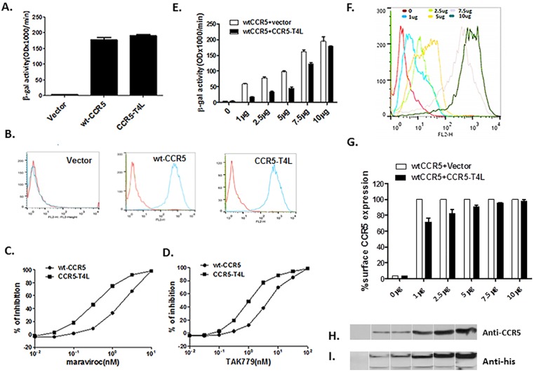
Fig 1. Recombinant CCR5-T4L acts as a HIV-1 co-receptor and down-regulates WT-CCR5 expression on the 3T3T4 cell surface.
A) HIV-1 co-receptor function was estimated using cell-cell fusion assays. 3T3.T4 cells were transfected with 15 μg pSC-CCR5-T4L, then infected with vaccinia virus encoding LacZ gene under T7 promotor and used as target cells. The target cells were mixed with HeLa effector cells, expressing BaL Env protein and bacteriophage T7 RNA polymerase. After incubation for 2.5 h at 37°C, the amount of β-galactosidase activity was measured. WT-CCR5 was used as a positive control. B) CCR5 expression on the cell surface was confirmed using FACS. The level of CCR5 expression on above target 3T3.T4 cells was measured by flow cytometry using either phycoerythrin (PE)-conjugated 2D7 or phycoerythrin (PE)-conjugated 3A9 monoclonal antibodies. C and D) CCR5-T4L was more sensitive to CCR5 agonists maraviroc and TAK-779 inhibition. Different concentrations of maraviroc or TAK-779 were used to pretreat target 3T3.T4 cells transfected with pSC-CCR5-T4L or pSC-CCR5 for 1 h at 37°C. After washing with PBS, cell-cell fusion was performed. E and F) CCR5-T4L inhibited WT-CCR5 by reducing its expression on the cell surface. Different amounts of CCR5-T4L or WT-CCR5 cDNA (0 μg、 1 μg、 2.5 μg、5 μg、 7.5 μg and 10 μg) were co-transfected into 3T3.T4 cells. The effect of R5 HIV-1 Env-mediated cell-cell fusion was examined (E) and the level of CCR5 expression on the cell surface was measured by flow cytometry using a PE-conjugated monoclonal antibody (2D7) (F). G) The inhibition by CCR5-T4L decreased as the amounts of CCR5-T4L and WT-CCR5 increased. CCR5 expression was measured using flow cytometry and a PE-conjugated monoclonal antibody (2D7). The averaged mean fluorescence values (MFVs) for CCR5 from three experiments are plotted as a bar diagram, where WT-CCR5 expression after transfection with WT-CCR5 plus empty vector is set at 100. H and I) The interaction between CCR5-T4L and WT-CCR5 was tested using co-immunoprecipitation. 3T3.T4 cells co-transfected with CCR5-T4L and WT-CCR5 were lysed with RIPA buffer. Lysates were prepared and immunoprecipitated with the CCR5 C-terminal antibody, fractionated by SDS-PAGE, and immunoblotted. Blots were probed with the N-terminal CCR5 antibody (H), stripped, and reprobed with the anti-6×His antibody (I). Following the primary antibody reaction, blots were washed and probed with the anti-mouse antibody conjugated to HRP. Blots were exposed to X-ray film after reaction with the HRP substrate.

