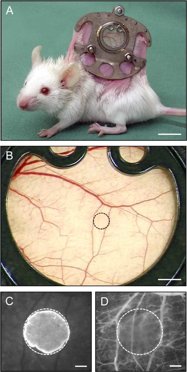
Fig 6. Dorsal skinfold chamber model for the in vivo analysis of tumor angiogenesis.

A: BALB/c mouse with a dorsal skinfold chamber (weight: ~2g). B: Observation window of a dorsal skinfold chamber directly after transplantation of a CT26 tumor cell spheroid (border marked by broken line). C, D: Intravital fluorescence microscopy of the tumor cell spheroid (border marked by broken line) in B. Because the cell nuclei of the spheroid were stained with the fluorescent dye Hoechst 33342 before transplantation, the implant can easily be differentiated from the non-stained surrounding host tissue of the chamber using ultraviolet light epi-illumination (C). Blue light epi-illumination of the identical region of interest as in C with contrast enhancement by intravascular staining of plasma with 5% FITC-labeled dextran 150,000 i.v. allows the visualization of the microvasculature surrounding the spheroid (D). Scale bars: A = 10mm; B = 1.4mm; C, D = 250μm.
