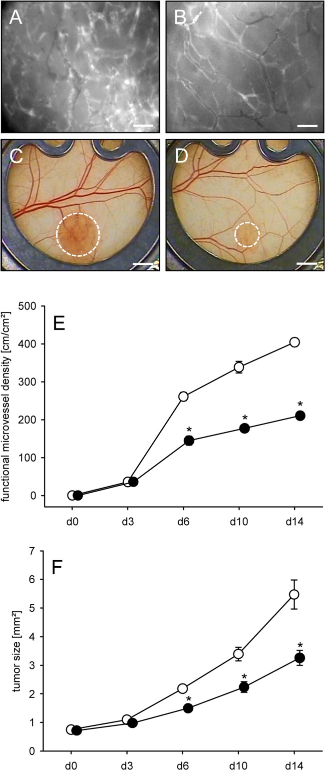
Fig 7. Geraniol action on tumor vascularization and growth.

A, B: Intravital fluorescence microscopic images of the newly developed microvascular network within CT26 tumors at day 14 after implantation into the dorsal skinfold chamber of a vehicle-treated control mouse (A) and a geraniol-treated animal (B). Blue light epi-illumination with contrast enhancement by 5% FITC-labeled dextran 150,000 i.v.. Scale bars: 50μm. C, D: Stereo microscopic images of CT26 tumors (borders marked by broken line) at day 14 after transplantation of spheroids into the dorsal skinfold chamber of a vehicle-treated (C) and a geraniol-treated animal (D). Scale bars: 1.4mm. E, F: Functional microvessel density (cm/cm2) (E) and size (mm2) (F) of CT26 tumors in dorsal skinfold chambers of vehicle-treated (white circles; n = 8) and geraniol-treated BALB/c mice (black circles; n = 8), as assessed by intravital fluorescence microscopy and computer-assisted off-line analysis. Means ± SEM. *P<0.05 vs. control.
