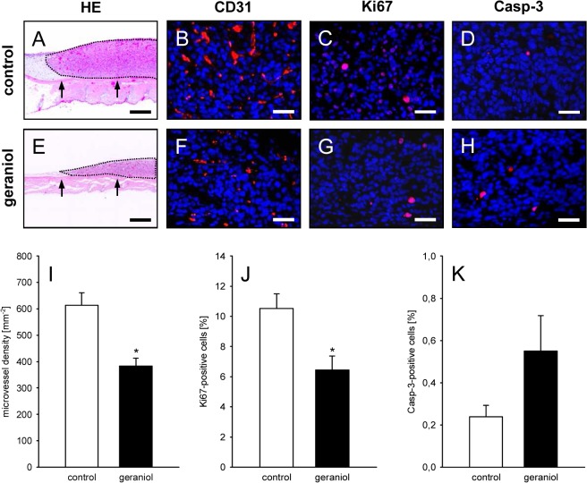
Fig 8. Histological and immunohistochemical analysis of tumors.
A, E: HE-stained cross sections of CT26 tumors (borders marked by dotted line) at day 14 after transplantation of tumor spheroids onto the striated muscle tissue (arrows) within the dorsal skinfold chamber of a vehicle-treated control mouse (A) and a geraniol-treated animal (E). Scale bars: 300μm. B, C, D, F, G, H: Immunohistochemical detection of CD31 (B, F, red), Ki67 (C, G, red) and Casp-3 (D, H, red) in CT26 tumors at day 14 after transplantation of tumor spheroids into the dorsal skinfold chamber of a vehicle-treated control mouse (B, C, D) and a geraniol-treated animal (F, G, H). Sections were stained with Hoechst 33342 to identify cell nuclei (blue). Scale bars: 40μm. I-K: Microvessel density (mm-2) (I), Ki67-positive cells (%) (J) and Casp-3-positive cells (%) (K) in CT26 tumors in dorsal skinfold chambers of vehicle-treated (white bars; n = 8) and geraniol-treated BALB/c mice (black bars; n = 8), as assessed by quantitative immunohistochemical analysis. Means ± SEM. *P<0.05 vs. control.

