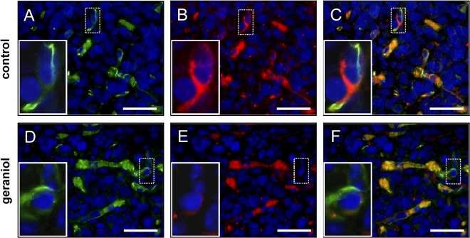
Fig 9. Immunohistochemical analysis of tumor microvessels.
Immunohistochemical detection of endothelial CD31 (A, D, green) and VEGFR-2 (B, E, red) of microvessels within CT26 tumors at day 14 after transplantation of tumor spheroids into the dorsal skinfold chamber of a vehicle-treated control mouse (A-C) and a geraniol-treated animal (D-F). Sections were stained with Hoechst 33342 to identify cell nuclei (blue). C and F are merges of A, B and D, E. Erythrocytes in the vessel lumina are unspecifically stained (C, F, orange color). Note that the endothelium of microvessels within the geraniol-treated tumor exhibits a markedly reduced expression of VEGFR-2 (E, insert = higher magnification of dotted ROI) when compared to those within the vehicle-treated control tumor (B, insert = higher magnification of dotted ROI). Scale bars: 35μm.

