
Fig 2.
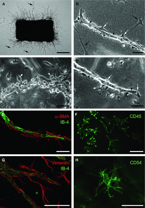
Aortic ring model of angiogenesis: cellular composition of angiogenic outgrowths. (A) Serum-free collagen gel culture of rat aorta photographed at day 6 (microvessels marked by arrows). (B) Microvessel composed of an inner core of endothelial cells (arrowheads) and surrounding pericytes (white arrows); black arrows indicate fibroblasts. (C) Linear cluster of macrophages (white arrowheads), with characteristically vacuolated and granular cytoplasm, at the root of an out-of-focus endothelial sprout (arrow). (D) Microvessel composed of endothelial cells (black arrowheads) and pericytes (white arrows) with surrounding macrophages (white arrowheads). (E) Confocal image of microvessel double stained with endothelial (isolectin-B4, IB-4) and pericyte (anti-α smooth muscle cell actin) markers. (F) Immunofluorescent image of macrophages stained for CD45. (G) Confocal image of aortic cultures double stained for endothelial cells (IB-4) and fibroblasts (vimentin). (H) Immunofluorescent image of dendritic cell stained for CD54 (ICAM). Magnification bars = 500 μm (A), 50 μm (B–H).