
Fig 6.
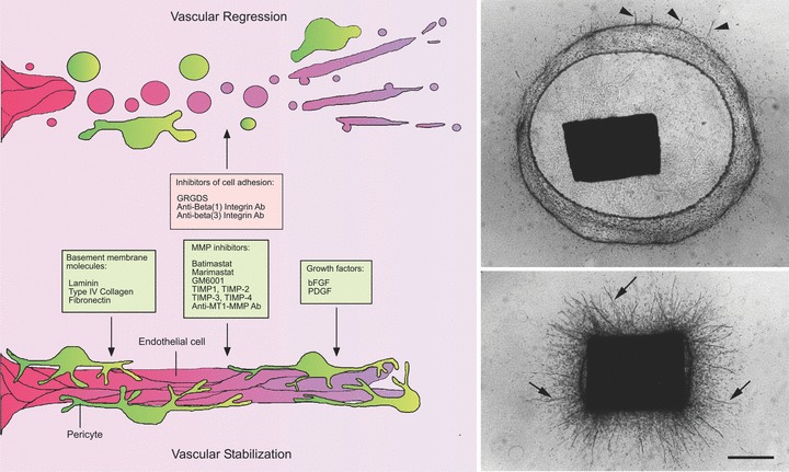
Vascular regression in the aortic ring model. Serum-free cultures of rat aorta can be used to study mechanisms of vascular regression following angiogenesis. Aorta-derived microvessels spontaneously regress after the first week of growth. Vascular regression is characterized by cell detachment from the ECM, vessel fragmentation and retraction of the remaining stumps. Molecules that interfere with cell attachment to the ECM such as RGD peptides and antibodies against β1 integrin or β3 integrins have the capacity to accelerate vascular regression. The anti-β1 integrin antibody induces vascular regression in collagen. The anti-β3 antibody has no effect in collagen but it synergizes with the anti-β1 integrin antibody to promote vascular regression in fibrin. Vessel survival can be extended by supplementing gels of interstitial collagen with basement membrane molecules and by blocking MMP activity with synthetic inhibitors, TIMPs or anti-MT1-MMP antibody. Vessel survival is also promoted by bFGF or PDGF. Photomicrographs show a control culture (upper panel) in an advanced stage of collagen lysis and vessel regression (residual short vessel stumps marked by arrowheads) and a parallel culture (lower panel) treated with the MMP inhibitor Marimastat to block collagen lysis and promote vascular survival (vessels marked by arrows). Magnification bar = 800 μm.