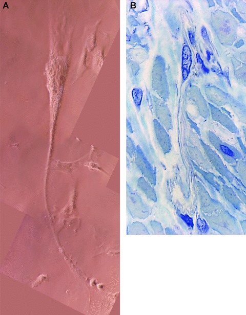
Fig 4.

(A) Human myometrium. Primary semi-confluent cell culture (day 4), phase contrast microscopy. Photographic reconstruction of an m-ICLC with a very long, moniliform cytoplasmic processes (arrows) emerging from cell body. Higher magnification (40×). (B) Human pregnant myometrium (39 weeks of gestation). Semi-thin sections (0.5- to –1-μm thick) of uterine muscular layer embedded in Epon resin and stained with toluidine blue. One may observe the very long process of m-ICLC squeezing between obliquely cut smooth muscle cells. Original magnification 100×.
