
Fig 7.
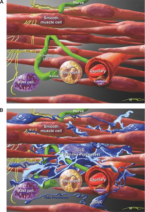
Artistic view of possible interconnectivity of m-ICLC in the uterine wall. (A) Myometrial background with smooth muscle cells, nerve fibers and some connective tissue cells. (B) Close proximity of m-ICLC processes with smooth muscle cells, unmyelinated nerves, capillaries, collagen fibres and immunoreactive cells. Scale bar – approximately 7 μm (erytrocyte’s diameter).