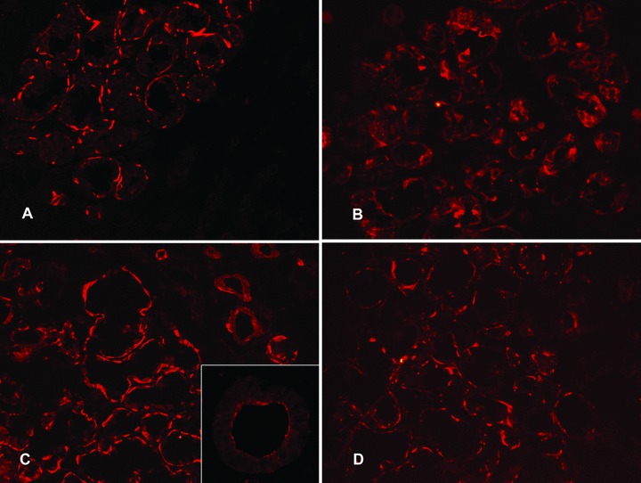
Figure 4.

Expression of 3β-hydroxysteroid dehydrogenase and 17β-hydroxysteroid dehydrogenase in labial salivary glands. In healthy controls, 3β-hydroxysteroid dehydrogenase (A) and 17β-hydroxysteroid dehydrogenase (C) stained the basal parts of the acinar cells leading to a narrow interrupted band-like staining. Insert in (C): In contrast, ductal staining was often characterized by a narrow band of apical staining. In labial salivary glands from patients with SS, 3β-HSD staining was more diffuse (B, compared to A) and the same pattern was seen in 17β-HSD staining (D, compared to C). Objective magnification ×200 (in A, B, C and D) or ×400 (in the insert).
