
Figure 5.
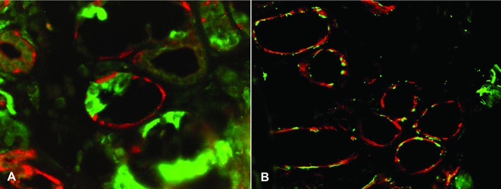
Hydroxysteroid dehydrogenases (HSD) were in the healthy labial salivary glands restricted to the mucous segments of the acini. (A) 17β-HSD (red colour) was stained together with lactoferrin (green colour), which is restricted to the serous demilunes. It was found that 17β-HSD only embraced the mucous segments of the acini, but did not embrace the seromucous half moons. (B) To exclude exclusive staining of myoepithelial cells, HSDs (17β-HSD in the panel, red colour) and myoepithelial cell marker smooth muscle actin showed very little overlap (which would produce yellow colour). Objective magnification ×200.