Figure 6.

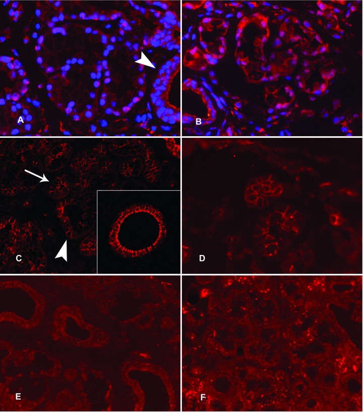
Expression of 5α-reductase and aromatase in labial salivary glands. In healthy labial salivary glands, 5α-reductase staining was strong in the nuclei of the acinar cells, whereas it had a more diffuse localization in the ductal epithelial cells (A, arrow head). This partial nuclear localization of 5α-reductase is very evident with nuclear DAPI counterstain (A). In contrast, aromatase immunoreactivity was polarized in the acinar cells so that it was expressed strongly on apical and lateral cell membrane domains (C, arrowhead) but was barely seen in the basal cell membrane (C, arrow) (C). Insert in (C): In salivary ducts aromatase was often seen also on the basal aspects of the ductal epithelial cells. In labial salivary glands of patients with SS, 5α-reductase staining was mostly cytoplasmic (B, compared to A). For the expression of aromatase in SS salivary glands, a similar expression was found as in healthy controls (D, compared to C). (E) and (F): The negative staining controls with normal, non-immune goat IgG, used instead of and at the same concentration as the specific primary antibodies, confirm the specificity of the stainings. Objective magnification ×200 in all panels and ×400 in the insert.
