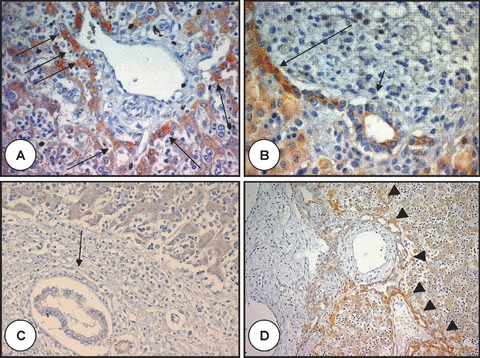
Figure 1.

Polyductin expression in foetal liver without and with ductal plate malformation. In (A), there is a portal tract at the stage of ductal plate with intense staining (arrows) of the hepatocytes at the limiting plate (anti-FP2, ×400); (B) shows a remodelling ductal plate structure with a gradient of staining intensity: the intensity is greater close to the periportal hepatocytes (long arrow) than close to the duct or in the remodelling bile duct (short arrow) (anti-FP2, ×400); (C) shows no staining of the remodelled bile duct (arrow) (anti-FP2, ×200). A baseline staining is present in the hepatocytes; (D) shows a liver of foetus with Meckel syndrome and ductal plate malformation with intense staining of the abnormal biliary structures (arrowheads) close to the periportal hepatocytes (anti-FP2, ×200).
