Figure 3.

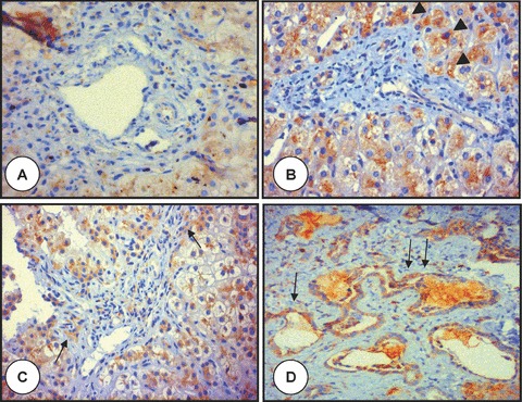
Polyductin expression in PIBD, AATD, PFIC I and Caroli’s disease. The microphotograph labelled with (A) shows a portal tract of a child with syndromatic paucity of the intrahepatic bile ducts (Alagille’s syndrome) showing practically baseline staining of the hepatocytes, but no staining in the portal area (anti-FP2, ×400); in (B) is shown the portal tract from an infant with alpha-1-antitrypsin deficiency with more intense FP-2 staining at level of the periportal area (arrowheads). The globules of accumulated alpha-1-antitrypsin are not seen because of the FP2 staining (anti-FP2, ×400); in (C) is shown the portal tract of an infant with progressive familiar intrahepatic cholestasis type I with moderate to intense staining of the biliary structures (arrows) and periportal hepatocytes (anti-FP2, ×400); (D) shows the liver tissue of a patient affected with Caroli’s disease. There is dilation of the bile ducts and intraluminal bulbar protrusions of the ductal wall with moderate to intense staining of the biliary structures (arrows) (anti-FP2, ×200).
