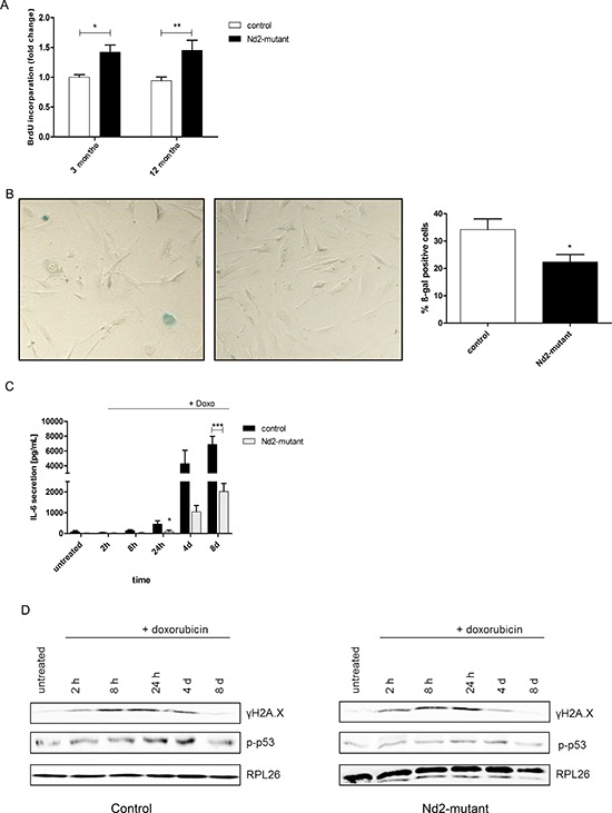
Figure 3. Characteristic features of cellular senescence in skin fibroblasts of Nd2-mutant mouse strain.

A. For primary skin fibroblasts of 3-month-old and 12-month-old mice, cell proliferation of Nd2-mutant or control mouse fibroblasts was analysed by BrdU incorporation assay. n = 3–13, *p < 0.05, **p < 0.01. B. Fibroblasts from 12-month-old mice were fixed and stained for SA-ß-gal 24 h after seeding. Representative picture of SA-β-gal-staining is shown (left panel). The number of positive blue cells was divided by the total number of counted cells resulting in the percentage of ß-gal-positive cells (right panel). n = 3, *p < 0.05. Data are expressed as mean ± SEM. C. Fibroblasts of 12-month-old Nd2-mutant and control mice were treated for 1 h with 250 nM doxorubicin and supernatants were collected and analysed for IL-6 concentrations by ELISA at indicated time points. n = 6–7, *p < 0.05 ***p < 0.001. Data expressed as mean ± SEM. D. Fibroblasts of 12-month-old Nd2-mutant and control mice were treated for 1 h with 250 nM doxorubicin and whole cell lysates were collected at the indicated days thereafter. Protein expression of γH2A.X and p-p53 was analysed by immunoblotting. Lysates were pooled from fibroblasts of three different mice. RPL26 was used as loading control.
