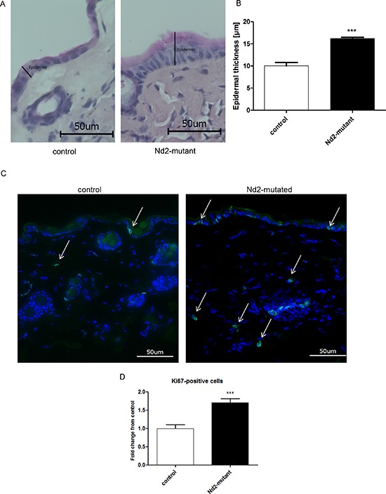
Figure 6. Measurement of epidermal thickness and Ki67 staining of the skin of Nd2-mutant mice.

A. Representative photomicrographs of H&E staining of skin sections from control (n = 4) and Nd2-mutant (n = 4) mice, aged 12 months. B. Quantification of the thickness of the epidermis in skin sections from control (n = 4) and Nd2-mutant (n = 4) mice using BZ II analyzer software. ***p < 0.001. Data expressed as mean ± SEM. C. Representative photomicrographs of Ki67 immunofluorescence staining (green) of skin sections from control (n = 5) and Nd2-mutant mice (n = 5), aged 12 month. DAPI was used for counterstaining of cell nuclei. D. Quantification of Ki67-positive cells in skin sections from control and Nd2-mutant mice using Image J software. ***p < 0.001. Data are expressed as mean ± SEM.
