
Figure 5. Effect of LINC00472 expression on breast cancer cell migration.
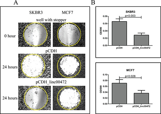
A. Microscopic views of cell migration before (0 hour) and after (24 hour) removal of stopper in SKBR3 and MCF7 cells transfected with pCDH or pCDH_LINC00472. Cells transfected with pCDH or pCDH_LINC00472 vectors formed monolayers in Oris 96-well plate and started to migrate to the exposed area after removing the stoppers in the well. Twenty-four hours later, the cells were fixed and stained with 0.1% crystal violet stain. The photomicrograph of the entire ‘wound’ area was taken under the IX71 inverted microscope with 4X objective lens. The representative wells were presented and the yellow circles indicated the areas previously occupied by stoppers. B. Measurements of absorbance after removal of stopper in SKBR3 and MCF7 cells transfected with pCDH or pCDH_LINC00472. With the detection mask, the absorbance at 590 nm wavelength of each well, which was directly proportional to the number of cells that migrated into the ‘wound’ area, was measured. The bar charts showed the average absorbance (y axis) from the wells with different cells after subtracting the background, the absorbance of the reference wells. Error bars represent SEM, n = 8. P values were determined by the Mann-Whitney U test.