
Figure 1. Physicochemical characterization and in vitro uptake of the cetuximab-IONPs.
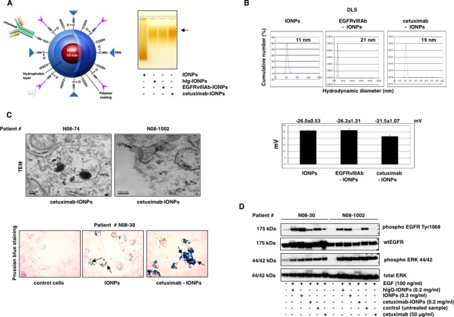
(A, left) Illustration of amphiphilic triblock copolymer-coated IONPs conjugated to cetuximab. (A, right) Confirmation of conjugation of IONPs to cetuximab, EGFRvIIIAb, and a human IgG by mobility shift (black arrow) in 1% agarose gel. (B, top) Dynamic light scattering (DLS) and hydrodynamic diameter of IONPs, cetuximab-IONPs, and EGFRvIIIAb-IONPs. (B, bottom) Zeta potential of IONPs, EGFRvIIIAb-IONPs, and cetuximab-IONPs. (C, top) Transmission electron microscopy (TEM) studies of cell binding and internalization of cetuximab-IONPs into lysosomes of human GBM neurospheres N08-74 and N08-1002 (magnification 10,000x). (C, bottom) Prussian blue staining of control (no treatment), IONPs, and cetuximab-IONPs internalized by human GBM neurospheres N08-30 (representative slides are shown). Neurospheres were allowed to attach to cell culture dish after treatment in neurobasal media. Nanoparticles are indicated by black arrows, magnification 40x. Cetuximab-IONPs showed maximal uptake. (D) Effect of cetuximab-IONPs on phosphorylation of EGFR after activation with EGF. Human GBM neurospheres N08-30 and N08-1002 were starved for 24 hs, pretreated with cetuximab-IONPs, IONPs, hIgG-IONPs, or cetuximab for 3 hs, followed by activation with 100 ng/ml EGF for 15 min. Western blotting with phospho-EGFR Y1068 antibody shows decreased activation of EGFR in the presence of cetuximab-IONPs. Total ERK 44/42 was used as an internal control.